 ECS is hosting a series of webinars presented by distinguished speakers this June. Join us! Speakers include Harry Atwater from the California Institute of Technology, Arumugam Manthiram from the University of Texas at Austin, and Paul Kenis from the University of Illinois at Urbana-Champaign. Topics include batteries, energy, carbon, and more. Considering attending? Learn more about what you can expect to hear about from our presenters! (more…)

Chilled Proteins and 3-D Images: The Cryo-Electron Microscopy Technology That Just Won a Nobel Prize
Posted on October 5, 2017 by Amanda StallerBy: Melanie Ohi, University of Michigan and Michael Cianfrocco, University of Michigan
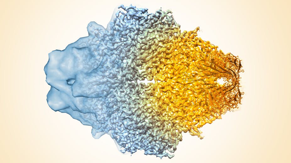
Cryo-electron microscopy resolution continues to improve. Veronica Falconieri, Sriram Subramaniam, National Cancer Institute, National Institutes of Health, CC BY-NC
Many people will never have heard of cryo-electron microscopy before the announcement that Jacques Dubochet, Joachim Frank and Richard Henderson had won the 2017 Nobel Prize in chemistry for their work developing this technology. So what is it, and why is it worthy of this honor?
Cryo-electron microscopy – or cryo-EM – is an imaging technology that allows scientists to obtain pictures of the biological “machines” that work inside our cells. Most amazingly, it can reconstruct individual snapshots into movie-like scenes that show how protein components of these biological machines move and interact with each other.
It’s like the difference between having a list of all of the individual parts of an engine versus being able to see the engine fully assembled and running. The parts list can tell you a lot, but there’s no replacement for seeing what you’re studying in action.
Posted in Announcements
![[Click to enlarge]](https://www.electrochem.org/wp-content/uploads/2015/10/PdNi_Fig10.jpg)
[Click to enlarge]
In a response to a recent call for photos, Galina Strukova sent us some great shots of the microworld of palladium-nickle alloy.
You’re looking at pictures of real objects of the palladium-nickel alloy, the size of the samples ranging from tens of micrometers to 1-2 millimeters.
They are produced via self-organization of nano-sized (several nanometer in diameter) wires growing on porous membranes under the action of electric current pulses. The authors have managed to isolate and photograph them by means of a modern electron microscope. They have described this 3D sculptures in scientific journals.
Such antenna-like samples are expected to find application in nanotechnology. Now we can produce such “sculptures” from various metals “by order,” examine them and admire their elegant forms.
However, it is still a riddle. Why does it so closely resemble plants and seashells? Does this mysterious self-organization have anything in common with formation of plant leaves, fungi, and seashells?
Read Strukova and Strukov’s previous installment, “The Beauty and Mystery of the Microworld.”
PS: Do you have interesting science photos you’d like us to share on the ECS Redcast Blog? Send your pictures and a short write-up to rob.gerth@electrochem.org. We’re always looking for great guest posts!
Posted in Guest Post
![[Click to enlarge]](https://www.electrochem.org/wp-content/uploads/2015/09/post-pic.png)
[Click to enlarge]
The beauty of these pictures is intriguing and fascinating by its asymmetric, exquisite and intricate pattern. What is it? Is it a product of a novel computer program or photographs of fine creations of nature? Neither statement is true. In fact, these are not pictures, but images of metal samples made with an electron microscope.
Only some color is added to the images to emphasize their resemblance to natural objects of our macroworld: seashells, jelly-fish, leaves of exotic plants. The size of the samples is from tens of micrometers to 1-2 millimeters. They are produced via self-organization of nano-sized (millionth of a millimeter) wires growing on porous membranes under the action of electric current pulses.
![[Click to enlarge]](https://www.electrochem.org/wp-content/uploads/2015/09/post-pic-2.png)
[Click to enlarge]
Besides, they have proved that the internal structure of this metallic “seashells” is a volumetric multilayer network woven by nano-sized wires. Such antenna-like samples are expected to find application in nanotechnology. Now we can produce such “sculptures” from various metals “by order”, examine them and admire their elegant forms and fascinating beauty. However, it is still a riddle. Why do they so closely resemble shells and leaves? Does this mysterious self-organization have anything in common with formation of plant leaves and seashells?
[1] J of Bionic Engineering 10 (2013) 368–376
[2] Materials Today 16 (2013) 98–99
[3] Materials Letters 128 (2014) 212-215
Posted in Guest Post
The microscope they developed produces x-ray images by scanning a sample while collecting various x-ray signals emerging from the sample.
Image: Brookhaven National Laboratory
Researchers have developed a new x-ray microscope that will provide scientists with the opportunity to image nanostructures and chemical reactions down to the nanometer.
The new class of x-ray microscope allows for nanoscale imagining like never before. This development brings researchers one step closer to the ultimate goal of nanometer resolution.
This from Brookhaven National Laboratory:
The microscope manipulates novel nanofocusing optics called multilayer Laue lenses (MLL) — incredibly precise lenses grown one atomic layer at a time — which produce a tiny x-ray beam that is currently about 10 nanometers in size. Focusing an x-ray beam to that level means being able to see the structures on that length scale, whether they are proteins in a biological sample, or the inner workings of a fuel cell catalyst.
Posted in Uncategorized
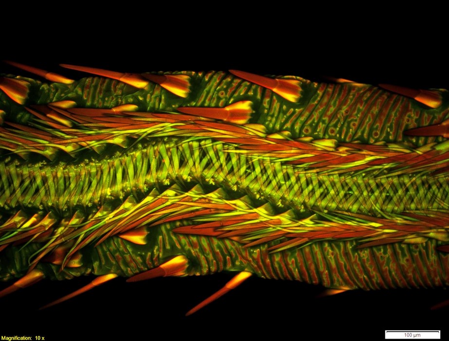
The Olympus BioScapes competition is held to celebrate the intersection of art and science.
Credit: Dr. Matthew S. Lehnert of Kent State University at Stark
We sometimes get so wrapped up in the technicality of science that we forget how beautiful it can be. Microscopy in particular provides us with the ability to see remarkable worlds that are otherwise invisible to the naked eye.
The Olympus BioScapes competition is held every year to help celebrate the intersection of art and science. Scientists from around the world submit their photos and videos of microscopy work to be judged “based on the science they depict, their beauty or impact, and the technical expertise involved in capturing them.”
Posted in Uncategorized
The image to your right may look like a fine art print of an ocean scene at night, but it’s actually just a close-up of some dried Glenlivet 162, or for those of you who aren’t avid alcohol connoisseurs – it’s simply a photo of whiskey.
Maybe “simple” is not the best word to describe the chemical process that takes place, but the discovery that whiskey can make these beautiful images had a humble beginning.
Professional artist and photographer Ernie Button started creating photos of the patterns formed after letting a drop or two of whiskey dry at the bottom of a glass, which resulted in these clear and rhythmic images.
Though he loved the aesthetic value, Button wanted to understand why the images looked the way they looked.
Posted in Education
Tagged alcohol, art, artist, beverage facts, chemical, chemistry, food science, Marangoni Effect, microscope, microscopy, photograph, photography, physics, science, whiskey
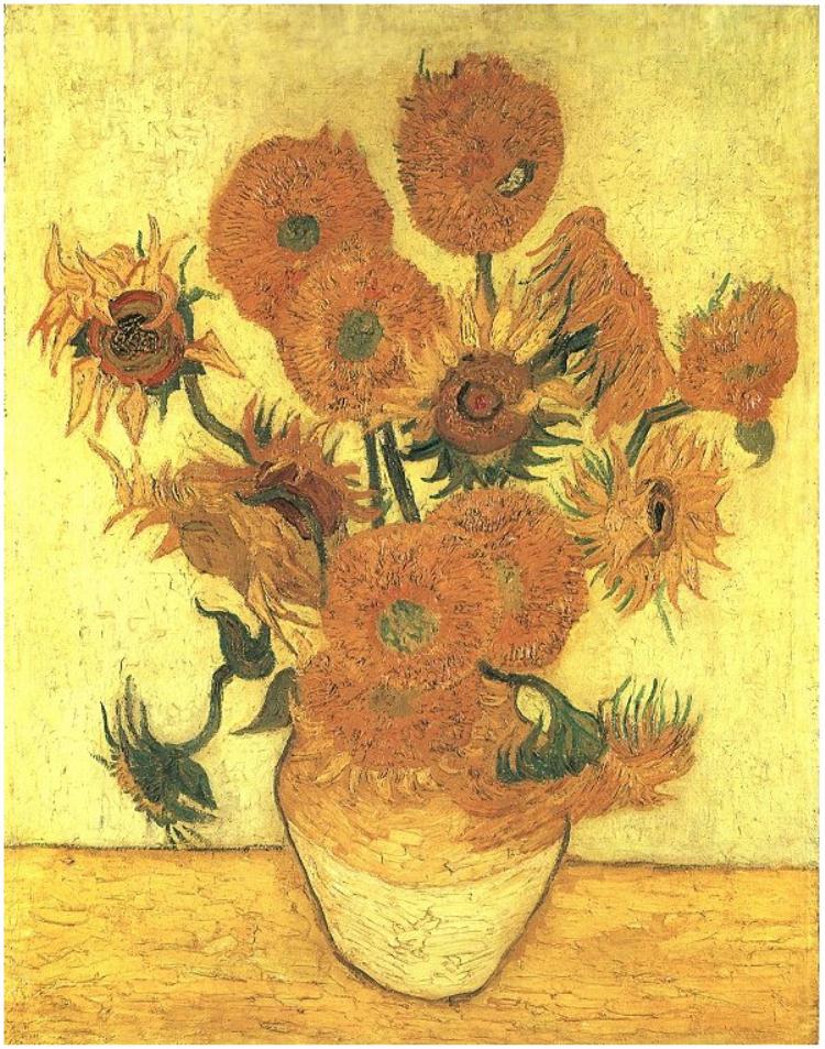
By examining paint segments from Van Gogh’s “Sunflowers,” experts believe preservation techniques could be improved.
Credit: Van Gogh Gallery
Electrochemical and solid state science transcend the limits of academic science to touch many of the things we come into contact with on a day-to-day basis, whether we know it or not. Most recently we’ve gotten a first-hand account of this at our Electrochemical Energy and Water Summit, where some of the brightest minds in electrochemical and solid state science came together to solve critical issues in global sanitation. Now, these sciences are even assisting in the preservation of culture.
Pin-sized painting samples from Vincent van Gogh’s “Sunflowers” painting have been extracted from the Van Gogh Museum and are now under the microscope at The University of Queensland’s Centre for Microscopy and Microanalysis (CMM).
UQ’s Professor John Drennan is leading the project, which aims to understand the aging characteristics of significant artworks in order to improve conservation techniques.
Posted in Uncategorized
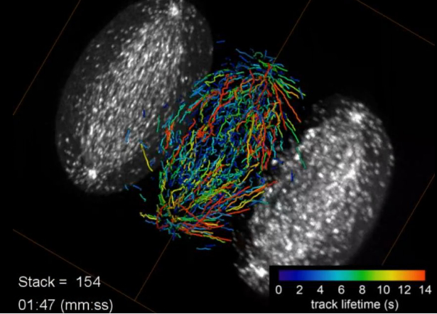
Growing microtubule endpoints and tracks are color coded by growth phase lifetime.
Credit: Betzig Lab, HHMI/Janelia Research Campus, Mimori-Kiyosue Lab, RIKEN Center for Developmental Biology
A new discovery out of Howard Hughes Medical Institute’s Janelia Research Campus is allowing biologists to see 3-D images of subcellular activity in real time.
They’re calling it lattice light sheet microscopy, and it’s providing yet another leap forward for light microscopy. The imaging platform was developed by Eric Betzig and colleagues in order to collect high-resolution images rapidly and minimize damage to cells.
Continue reading to check out the amazing video that shows the five different stages during the division of a HeLa cell as visualized by the lattice light sheet microscope.
Posted in Uncategorized
Tagged Bessel, Betzig, cells, chemistry, high resolution, lattice, lattice light sheet, light, microscope, microscopy, Nobel Prize, subcellular
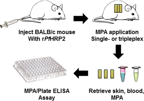
The researchers optimized their device so it could capture two biomarkers for the malaria parasite, Plasmodium falciparum, which kills more than 1 million people every year.
One day, there may be no poking or prodding for a blood sample when you go to the doctor.
In the American Chemical Society journal Analytic Chemistry, researchers report that they have developed a patch that can detect malaria proteins in live mice, and believe that this same technology could be adapted for use in humans to diagnose other diseases.
Along with being generally painful, drawing blood requires trained personnel and expensive lab equipment and facilities for analysis. With the development of this patch, scientists believe they will be able to overcome these obstacles in the near future, thereby providing better health care for resource-limited patients.
This from the American Chemical Society:
Scientists have been trying to address these hurdles by developing diagnostic patches that are covered on one side with thousands of microscopic, hollow needles that can sample fluid in the skin. But so far, these devices have only been able to test for one compound at a time. However, many diseases can be diagnosed more reliably by detecting multiple biomarkers. Corrie’s team wanted to design a new patch that could meet this need.
Posted in Uncategorized